Introduction
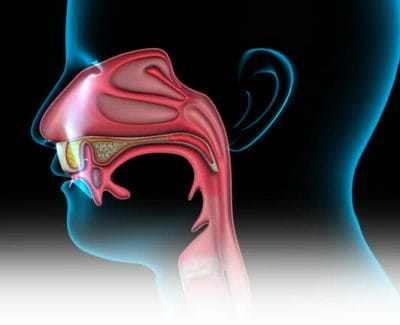
The term “skull base” refers to the bottom of the skull. It is a plate of bone upon which the brain sits. The skull base separates the brain from the remaining anatomic structures in the head and neck. These structures include the nasal cavity, sinuses, eyes, ears, and upper part of the throat. The skull base contains many openings called foramen. Nerves and blood vessels travel through the foramen to reach other parts of the head, neck and body. Infections and tumors of the sinuses can impact the skull base, the nerves coming out of the brain and the brain itself.
Anatomy
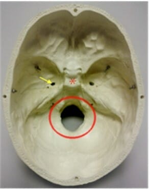
The skull base is made up of several different bones that are fused (joined) together. These fused bones look much like a bowl with holes in the bottom (See Figure 1). The large hole in the skull base is where the spinal cord leaves the brain and travels through the skull to the neck and back. This opening is known as the foramen magnum. The carotid artery traverses from the neck through the carotid foramen to help supply blood to the brain. The nerves that travel through the foramen ovale allow you to chew your food and give sensation to the lower part of your face.
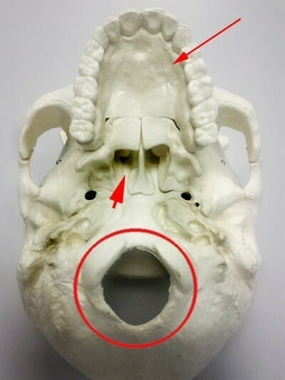
When flipped over, the skull base can be viewed from the bottom side (See Figure 2). Although the view is very different, the holes and crevices can again be seen. What is different is that other important structures can also be seen. These include the upper jaw and palate as well as the openings in the back of the nose (choanae).
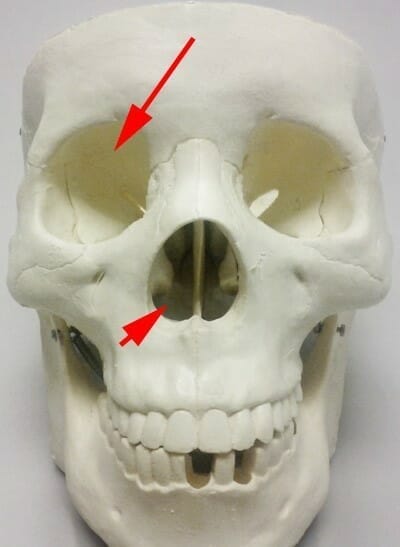
When we view the front of the skull, the image is more familiar (See Figure 3). This is the framework of the human face. The eye sockets (orbits) are present, as is the nasal cavity.
Importance of the Skull Base
There are a number of skull base disorders for which you may be referred to an Ear, Nose and Throat surgeon that specializes in this area (Rhinologists). Because the sinuses are located just below the skull base, Rhinologists are well-equipped to treat these conditions.
The fluid that surrounds the brain is called cerebrospinal fluid (CSF). In rare cases, CSF can leak through the skull base, into the sinuses and nose (See CSF Leaks). A CSF leak can occur after trauma, such as a motor vehicle accident, or can occur spontaneously.
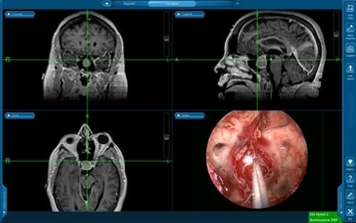
In addition, many types of tumors can occur in the area of the skull base. These including nasal, sinus, orbital and pituitary tumors. The pituitary gland sits in the middle of the skull base and is directly above and behind the sphenoid sinus. This is an area that rhinologists are able to access for surgery in a minimally invasive manner (See Figure 4).
Surgery of the Skull Base
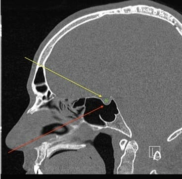
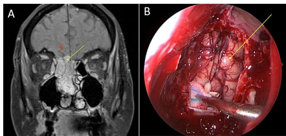
The skull base can be reached during surgery through a number of different “approaches” (or routes, See Figures 5 and 6). Traditional approaches involve creating incisions on the skin. These incisions can be on the face, on the scalp, behind the ear or on the neck under the jaw bone. While it may be necessary to use incisions to reach the skull base in some cases, in many situations the skull base can be reached with an endoscope (See Nasal Endoscopy). Endoscopic skull base surgery is considered a “minimally invasive” way of performing surgery. This method is often preferred, as it may involve fewer risks, lower complication rates and faster recovery than traditional approaches, with as good or better results. However, the best approach for each patient depends on a number of factors.
Is Endoscopic Skull Base Surgery Right for Me?
The decision to perform endoscopic skull base surgery depends upon many factors. Most importantly, the surgeon or surgeons must decide if the problem can be treated completely through the endoscopic approach. This depends on the type of tumor, the location of the tumor or defect, involvement of surrounding structures, and patient factors. If you have a sinus tumor, sinus cancer, CSF leak, pituitary tumor or other condition that involves the skull base, you should discuss with your surgeon what is best for you.
Copyright © 2020 by the American Rhinologic Society

