
Understanding the Paranasal Sinuses
The paranasal sinuses, often referred to as “the sinuses,” are hollow spaces found within the facial bone called the frontal bone. Each sinus is named after the bone it’s located in. Here’s a brief look at each:
- Maxillary Sinuses: These are found on each side of the face within the cheekbone.
- Ethmoid Sinuses: They form a honeycomb-like structure of small sinuses between the eyes.
- Frontal Sinuses: One on each side within the forehead bone.
- Sphenoid Sinuses: One on each side behind the ethmoid sinuses.


The Nasal Cavity
Inside the nose, the pink membranes lining the sinuses are called mucosa. They produce mucus, which helps keep the sinuses moist. The inside of the nose is called the nasal cavity. Along the sides of the nasal cavity are bone shelves called turbinates. These turbinates, covered by mucosa which helps humidify the air we breathe. There are typically three turbinates on each side (inferior, middle, and superior).
Most sinuses drain into a common pathway just beyond the middle turbinate, known as the osteomeatal complex. Air flows between the turbinates and the nasal septum, which separates the two sides of the nose. Eventually, both air and mucus are directed to the nasopharynx, where the nose meets the throat. From there, air enters the windpipe and lungs, while mucus is swallowed.
Additional Related Structures
Near the sinuses, there are several crucial structures:
- Tear Duct (Nasolacrimal Duct): It drains tears from the inner corner of the eye into the nasal cavity.
- Eustachian Tube: This tube helps equalize pressure in the ears and opens into the nasopharynx.
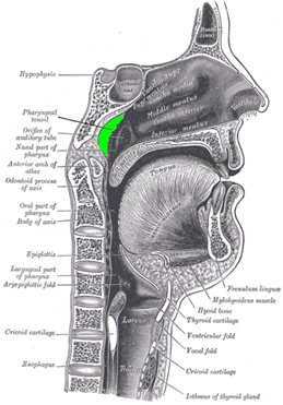
- Adenoids: These are tonsil-like tissues found at the top of the nasopharynx. While they can be large in children, they usually shrink during puberty. (Figure 4)
Understanding the surrounding anatomy is important for sinus health and surgical planning. For detailed insight into the bones and structures that support the sinuses, refer to skull base anatomy. This helps explain how sinus infections or procedures may affect adjacent areas like the eyes, ears, and brain.
Copyright © 2020 by the American Rhinologic Society